All About Mammograms! Procedures, Techniques & More
If you're a woman nearing 40 years of age, then it's high time to begin thinking about scheduling your first mammogram! Mammography, a diagnostic radiology medical imaging technique, is a crucial tool for the early detection of breast cancer.
But what is a mammogram, and what is it used to determine about breast health?
Table of Contents
- What is a mammogram anyways?
- Types of mammograms
- What should I expect the day of my mammogram?
- Interpreting your mammogram
- BI-RADS classification: Breast Imaging Reporting and Data System
- Mammography benefits, risks and limitations
- Conclusion
What is a mammogram anyways?
Mammography is a critical practice in your breast cancer detection and prevention toolkit that offers a window into the health of breast tissue. As a woman's risk of breast cancer increases with age, understanding mammograms becomes vital.
Mammograms, in short, are x-ray images of the breast used for early detection of breast cancer.
There are two types of mammogram procedures: screening mammograms for asymptomatic women and diagnostic mammograms for those with symptoms or abnormalities. The procedure itself involves compressing the breast between two plates to spread out the tissue for a clearer image.
While traditional film mammography has dominated the breast cancer imaging practice over the decades, in more recent years, digital mammography has largely taken its place as common practice.
This is generally because its output yields superior image quality, and is easier to store and transmit too!
Types of mammograms
2D mammograms or 'traditional mammograms' produce flat images of the breast tissue:
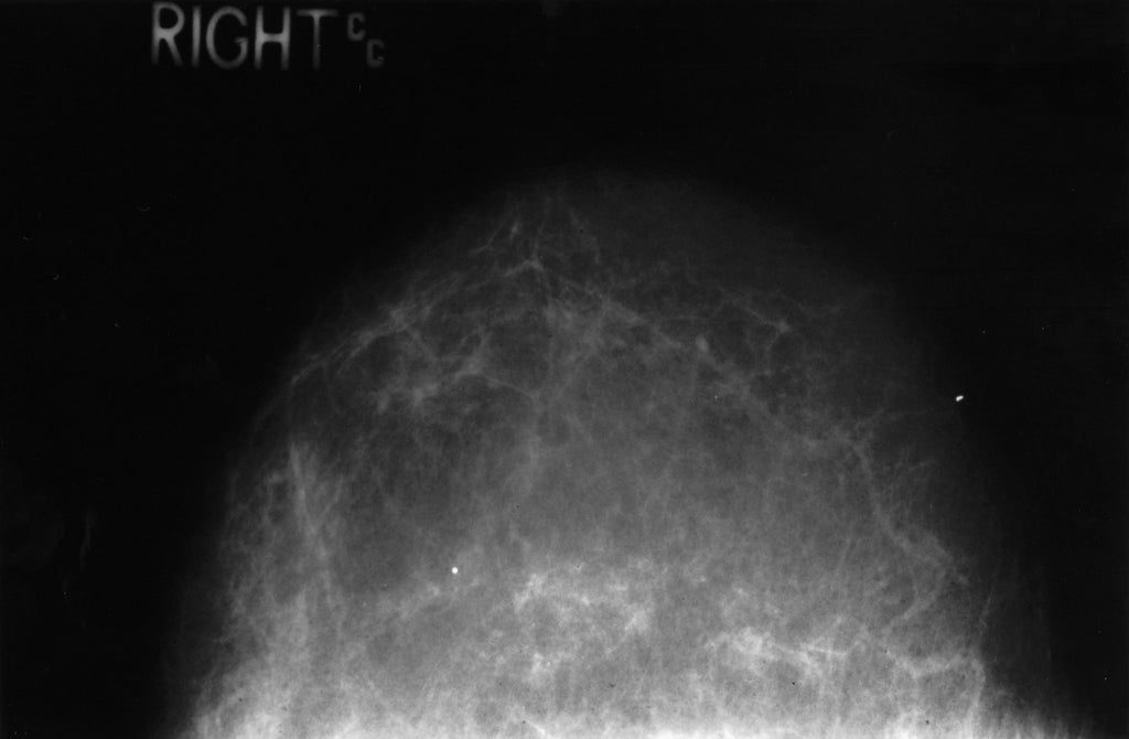
2D Film Mammogram of a normal, health breast. Courtesy of the National Cancer Institute
3D mammograms, also known as breast tomosynthesis, is an advanced technology that creates a three-dimensional image, reducing overlapping tissue and improving accuracy.
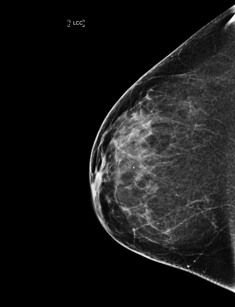
3D Breast Tomosynethesis of a normal, healthy breast. Courtesy of the National Cancer Institute.
A third type of mammography is known as contrast-enhanced mammography (CEM). It uses a contrast agent or dye containing iodine alongside the standard 3D mammogram to better highlight abnormal blood vessels. Within this practice, imaging can be performed through high-energy (HE), low-energy (LE), and dual-energy subtraction (DES).
Court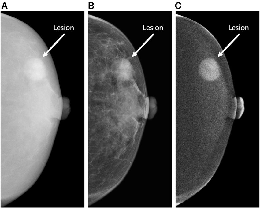
Contrast enhanced mammography can significantly enhance the detection of breast cancer in dense tissue.
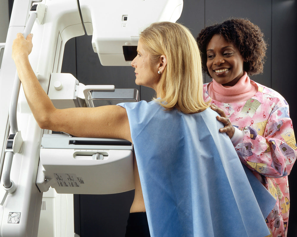
What should I expect the day of my mammogram?
We've outlined the basics of this common breast imaging procedure, but preparing for the procedure starts before you arrive.
Before the mammogram, your radiology team will advise that you avoid using the following to prevent interference with the images:
- Deodorant
- Lotions
- Powders
During the procedure, the technologist positions the breast on the machine and applies compression. This may cause some slight discomfort but it is necessary for accurate imaging!
Keep in mind that multiple images will be taken from different angles to capture the entire breast tissue.
Interpreting your mammogram
Your radiologists will interpret your mammograms by looking for any signs of breast cancer such as masses, calcifications, or architectural distortions.
The white components of the mammogram's output are densities in the breast tissue. These generally make it more difficult to detect tumors.
Breast calcifications
These are tiny deposits of calcium that can appear in breast tissue, and are typically detected in mammography. They typically present as tiny white specks on your mammogram.
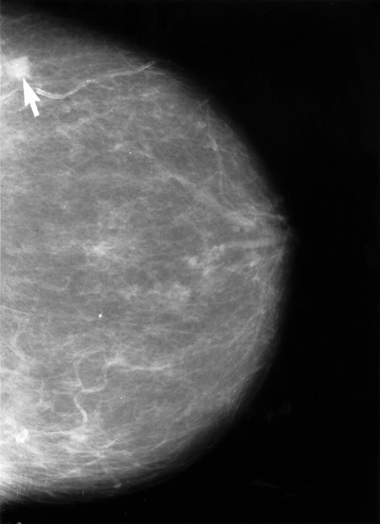
Microcalcifications are tiny, and are often benign (non-cancerous). However, they can present as an early sign of breast cancer.
Macrocalcifications tend to be quite larger, and are usually benign. They typically onset with age, or result from inflammation or other generally benign breast conditions.
In either case, your radiologist may schedule a follow up evaluation, just to be safe.
Tumors
Tumors are abnormal growths of tissue that can develop anywhere in the body, including the breast.
Benign breast tumors are non-cancerous, and typically don't spread beyond the breast tissue. Common types of benign breast tumors include fibroadenomas, cysts, and papillomas.
Malignant breast tumors will be routinely identified as breast cancer. They typically occur when abnormal cells in breast tissue grow uncontrollably and spread to the surrounding areas.
Breast cancer can originate in the milk ducts, also known as ductal carcinoma, or the milk producing lobules, also known as lobular carcinoma, of the breast.
Breast density and mammograms
Dense breast tissue is common. If you have dense breast tissue, it could increase your risk of breast cancer while making it more difficult to interpret growths within mammograms.
If you have dense breast tissue and are considered at-risk of developing breast cancer, your radiologist may recommend additional types of screenings, like MRIs or ultrasounds. This is to improve their chances of successfully detecting and diagnosing cancerous growths.
With all of these types of growths that may present in a mammogram, it's important to have a standardized classification system for accurate pathology. The BI-RADS classification system provides just that.
BI-RADS Classification: Breast Imaging Reporting and Data System
BI-RADS, the breast imaging reporting and data system, was established in 1993 by the American College of Radiology (ACR) to categorize mammogram findings.
It plays a crucial role in helping to standardize the communication between radiologists and referring physicians, which helps to guide patient management and ensure quality assurance in the field of breast imaging.
BI-RADS categorizes mammogram findings on a scale of 0-6; each category has its own specific management recommendations:
- BI-RADS 0: Incomplete assessment, additional imaging evaluation needed.
- BI-RADS 1: Negative (no significant abnormality detected).
- BI-RADS 2: Benign finding (e.g., cysts, calcifications, or fibroadenomas).
- BI-RADS 3: Probably benign (follow-up recommended, usually with short-term imaging).
- BI-RADS 4: Suspicious abnormality (suspicious for malignancy, further evaluation and/or biopsy recommended).
- BI-RADS 5: Highly suggestive of malignancy (appropriate action, such as biopsy, warranted).
- BI-RADS 6: Known biopsy-proven malignancy (previously confirmed breast cancer).
Benefits, risks and limitations
Getting a mammogram is a critical choice in monitoring one's own health to promote a long, healthy life.
But did you know that treatment for breast cancer is also most effective if it's caught in its earliest stages too?
In fact, early detection and treatment of breast cancer is key to preventing its spread throughout the body. If an early, localized detection is graded as BI-RADS 3 or 4, the 5-year survival rate ranges as high as 99% according to a 2021 Korean Surgical Society study.
For this reason, women can begin mammogram screenings annually as early as 40 years of age. At 45 years of age, it's recommended that annual mammograms begin across the board.
Reduced mortality rates is a major benefit of early detection mammography. From a risk perspective, mammograms are widely considered safe and their benefits are widely regarded as outweighing their risks. The radiation dosage is low, the facilities and equipment are regulated, and the procedures are optimized for safety.
While mammograms can yield false-positive or false-negative results, follow up appointments are common and lead to positive health outcomes.
Conclusion
In conclusion, mammograms are a cornerstone of breast cancer screening and early detection, and offer women the opportunity for timely intervention and improved outcomes.
Understanding the procedures, techniques, interpretation, benefits, risks, and BI-RADS classification empowers individuals to make informed decisions about their breast health!
By prioritizing regular mammograms and staying informed, we can work towards reducing the impact of breast cancer on women's lives.
Have any questions or would like to schedule an appointment with one of our Southern California radiologists? Give us a call or schedule an appointment today!